Unraveling the Mysteries of Brain Activity: A New Frontier in Brain Imaging
The human brain is a complex and intricate organ, consisting of billions of neurons that communicate with each other through electrical signals. Understanding how these neurons work together to enable our thoughts, emotions, and actions has long been a subject of fascination for scientists. One method that has gained significant attention in recent years is resting-state functional magnetic resonance imaging (rsfMRI), which measures brain activity by observing changes in blood flow to different parts of the brain. However, rsfMRI does not directly explain how these blood flow changes relate to neural activities.
To address this knowledge gap, a team of researchers led by Nanyin Zhang, a renowned expert in brain imaging and biomedical engineering at Penn State University, set out to investigate the relationship between rsfMRI signals and neural activity. Their findings, published recently in eLife, have shed new light on the complex dynamics of brain activity and challenge our current understanding of resting-state brain networks.
How Does rsfMRI Work?
Resting-state functional magnetic resonance imaging (rsfMRI) is a non-invasive technique that allows scientists to study how different parts of the brain work together. This method measures brain activity by observing changes in blood flow to different regions of the brain. The idea behind rsfMRI is simple: when a region of the brain is active, it consumes more oxygen and produces more carbon dioxide, which in turn increases the concentration of carbon dioxide in the bloodstream. This increased carbon dioxide triggers a response from the blood vessels, causing them to dilate or constrict, depending on the level of activity.
By analyzing these changes in blood flow, researchers can reconstruct maps of brain activity, revealing coordinated patterns of neural connectivity known as resting-state brain networks (RSNs). These RSNs are thought to play a crucial role in regulating various brain functions, including perception, attention, and memory.
However, despite the widespread use of rsfMRI in many research fields, there is still much we do not know about its underlying mechanisms. The current understanding of RSNs and rsfMRI signals is based on the assumption that the electrophysiology signal underlies most of the rsfMRI signal. However, this assumption has been challenged by recent studies, which have found discrepancies between the spatial and temporal relationships between rsfMRI signals and neural activity.
Addressing the Limitations of rsfMRI
To investigate these limitations, Zhang’s team used a novel technique that combines simultaneous recordings of rsfMRI and electrophysiology signals. This approach allows researchers to measure both the blood flow changes associated with neural activity and the actual neural activity itself. By analyzing these two signals in tandem, the team aimed to elucidate exactly how spontaneous blood flow changes in the brain are related to neural activities.
The Results: A Paradoxical Discovery
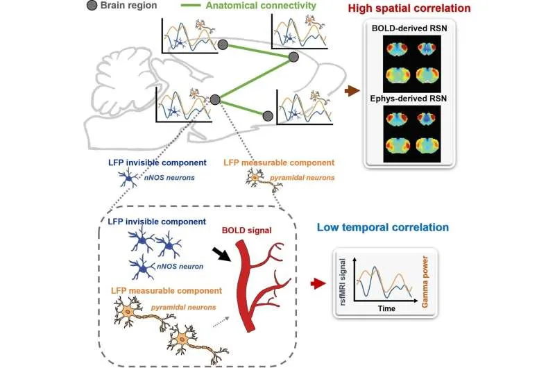
After months of data collection and analysis, Zhang’s team made a surprising discovery. The brain-wide RSN connectivity patterns revealed by rsfMRI signals could be recapitulated by electrophysiology signals. However, these two types of signals did not align well over time, suggesting that there are electrophysiology “invisible signals” contributing to the rsfMRI signal.
These findings imply that our current understanding of the neural basis of rsfMRI signals and RSNs might be incomplete or inaccurate. The possibility that a major source of the rsfMRI signal could originate from an electrophysiology-invisible component challenges the conventional view that the electrophysiology signal underlies most of the rsfMRI signal.
Implications for Brain Imaging
The implications of Zhang’s findings are far-reaching and profound. If our current understanding of RSNs and rsfMRI signals is incomplete or inaccurate, it could have significant consequences for brain imaging research. The possibility that there are electrophysiology-invisible components contributing to the rsfMRI signal suggests that we might need to revisit our assumptions about the neural basis of brain function.
Furthermore, the discovery of these invisible signals opens up new avenues for investigation into the neural mechanisms of brain activity. By exploring these hidden patterns, researchers may uncover new insights into the workings of the human brain and potentially develop more accurate methods for imaging brain activity.
Translating Animal Studies to Humans
The findings in this study have high translational value to human rsfMRI studies. Since the neural mechanism of rsfMRI in animals is likely to be similar to that in humans, results from animal models can provide valuable insights into the human brain. The use of animal models also allows researchers to control for variables that cannot be controlled in human studies, such as age, sex, and health status.
Conclusion
The discovery of electrophysiology-invisible components contributing to rsfMRI signals represents a significant breakthrough in our understanding of brain activity. Zhang’s team has challenged our current assumptions about the neural basis of RSNs and rsfMRI signals, opening up new avenues for investigation into the complex dynamics of brain function.
As we continue to explore the mysteries of brain activity, it is essential that we remain open-minded and willing to challenge our existing knowledge. By embracing these new findings and exploring their implications, researchers may uncover new insights into the workings of the human brain and develop more accurate methods for imaging brain activity.
The Future of Brain Imaging
The study published by Zhang’s team represents just the beginning of a new frontier in brain imaging research. As we continue to push the boundaries of what is possible with rsfMRI and other techniques, we may uncover even more secrets about the human brain.
The possibilities are endless, from exploring the neural mechanisms of neurological disorders to developing more accurate methods for diagnosing and treating brain injuries. Whatever the future holds, one thing is certain: the discovery of electrophysiology-invisible components contributing to rsfMRI signals has opened up new avenues for investigation into the complex dynamics of brain function.
And so, as we embark on this exciting journey of discovery, it is essential that we remain curious, open-minded, and willing to challenge our existing knowledge. For it is only by embracing these new findings and exploring their implications that we may uncover the secrets of the human brain and unlock its full potential.

0 Comments